
Unknown Choroidal Tumor Case 5
|
Subject: Unknown
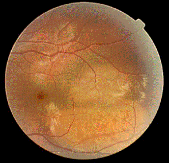
Date: August 19th, 2001 From: Barrett Haik MD We have a interesting case and would like to get your comments. Case This 20 year old male presented with a 4 week history of decreased vision OS. The referring doctor noted a mass temporal to the macula with surrounding fluid. What will be your approach in this case?
|
|
Responses From ECN Members
Tue, 21 Aug 2001 06:30:15 +0930
From: James Muecke, MD
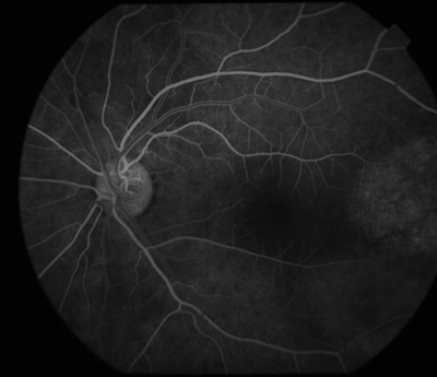
The ICG series is consistent with a solitary choroidal hemangioma with early hyperfluorescence and late washout, however it would be nice to see the very early images looking for the characteristic lacy pattern of fluorescence. I would not perform a biopsy, but US would be nice to see, although not diagnostic. There is no point in performing CT/MRI. These tumours respond very well to low dose lens-sparing external beam radiotherapy (3000 cGy in 200 cGy fractions) with reabsorption of the SRF within a month and slow gradual shrinkage of the tumour. Laser will not destroy the tumour and the SRF usually returns.
Best wishes,
James Muecke,
MD - Australia
Tue, 21 Aug 2001 06:30:15 +0930
From: James Muecke, MD
The ICG series is consistent with a solitary choroidal hemangioma with early hyperfluorescence and late washout, however it would be nice to see the very early images looking for the characteristic lacy pattern of fluorescence. I would not perform a biopsy, but US would be nice to see, although not diagnostic. There is no point in performing CT/MRI. These tumours respond very well to low dose lens-sparing external beam radiotherapy (3000 cGy in 200 cGy fractions) with reabsorption of the SRF within a month and slow gradual shrinkage of the tumour. Laser will not destroy the tumour and the SRF usually returns.
Best wishes,
James Muecke,
MD - Australia
Tue, 21 Aug 2001 17:18:28
From: Ms. Cathy Dibernardo
Standardized Echography (combined use of B-scan and Standardized A-scan) would be useful to secure the diagnosis. Typically, hemangioma exhibit high internal reflectivity, regular internal structure and no vascularity on Standardized A-scan. Unlike melanoma where the echographic features are low internal reflectivity, regular internal structure and mild, moderate or marked vascularity, or metastatic lesions that have an irregular shape, structure and reflectivity.
Baltimore - USA
Wed, 22 Aug 2001 18:47:04 +0300
From: Ihab S. Othman, MD
I agree with Dr. Muecke with the presumptive diagnosis of choroidal hemangioma based on ICG findings. However, I would like to address two points that might be controversial. First, the usefulness of MRI in diagnosis of hemangiomas of the choroid. As a diagnostic tool, MRI findings in such cases differ from other intraocular tumor being isointense to slightly hyperintense on T1 weighted image, and isointense to the vitreous on T2 weighted images with marked enhancement to gadolinium. Those MRI features are important in differentiating choroidal hemangioma from intraocular melanoma and metastases. On the other hand, the choroidal hemangioma is showing mild activity with minimal exudative retinal detachment.
Argon laser photocoagulation has been useful to dry out SRF, with a success rate of about 40% at 5 years interval as reported by Shields et al in the eighties. We have reported the use of diode laser thermotherapy in juxtafoveal choroidal hemangioma with great success (Othmane et al, Archives of Ophthalmology. 1999). Photodynamic therapy has recently been introduced to avoid the complications of a positive visual scotoma associated with TTT. We use external beam radiotherapy with a dose of 3000 cGy for highly active tumors with an extensive subretinal fluid (this is not the case). Doses as low as 2000 cGy may induce complete tumor inactivation in such a small tumor with minimal RD. (Alberti W. Radiotherapy of choroidal hemangioma. Int J. Radiat Oncol Biol Phys. 12: 122-123, 1986)
Sincerely,
Ihab S Othman,MD - Cairo , Egypt
Wed, 22 Aug 2001 11:40:16
From: Laurence Desjardins, MD
Clinically the lesion does not look very much like an hemangioma but more like amelanotic melanoma or metastasis. I would suggest before the biopsy, a general work up and transillumination.
Sincerely,
Laurence Desjardins, MD - Paris, France
From: Laurence Desjardins, MD
Clinically the lesion does not look very much like an hemangioma but more like amelanotic melanoma or metastasis. I would suggest before the biopsy, a general work up and transillumination.
Sincerely,
Laurence Desjardins, MD - Paris, France
Thurs, 23 Aug 2001 18.25
From: James Muecke, MD
I agree it does not look classical for a hemangioma, but they can certainly look like that. The ICG findings are classical however, and completely different from a metastasis or choroidal melanoma. A systemic work is always sensible.
In my opinion, the problem with using a lower dose (2000 cGy) is that the subretinal fluid reabsorbs but tumour bulk is not destroyed. If this happens, the patient may get persistent trouble with distorted vision. We have getting much better results with the higher dose. The visual results after radiation therapy are also far superior than with laser treatment for parafoveal hemangiomata with subretinal fluid extending into the fovea, I have been getting return to 6/6 within one month and there is no reaccumulation of SRF. External beam radiation therapy appears to be a permanent solution! Lastly, PDT is extraordinarily expensive for patients in Australia!!
Best wishes,
James Muecke, MD
Thu, 23 Aug 2001 16:37
From: Mr. Mark Evans
In my opinion, a good echographer should be able to differentiate between a choroidal hemangioma and a melanoma. In the COMS, our abilty to ultrasonographically diagnose choroidal melanoma was well above 90%.
Sincerely,
Mark Evans - Oregon, USA
From: Mr. Mark Evans
In my opinion, a good echographer should be able to differentiate between a choroidal hemangioma and a melanoma. In the COMS, our abilty to ultrasonographically diagnose choroidal melanoma was well above 90%.
Sincerely,
Mark Evans - Oregon, USA
Tuesday, 17th September 10:45
From: Maria Antonietta Blasi
The clinical and ICG findings are consistent with a diagnosis of choroidal hemangioma, but I think that an echography detecting high internal reflectivity would be useful. this hemangioma looks symptomatic and needs to be treated. TTT seems to be a good option.
From: Maria Antonietta Blasi
The clinical and ICG findings are consistent with a diagnosis of choroidal hemangioma, but I think that an echography detecting high internal reflectivity would be useful. this hemangioma looks symptomatic and needs to be treated. TTT seems to be a good option.
DISCLAIMER: Postings on The ECN Mailing List are strictly the opinions of the authors. The ECN and its sponsors assume no responsibility for the accuracy of the information, nor do they assure the safety or effectiveness of any clinical recommendations in these postings.
Receive the latest news and opportunities from The Eye Cancer Foundation. Please fill out the form below.
Receive the latest news and opportunities from The Eye Cancer Foundation. Please fill out the form below.
