Timing and the diagnosis of Uveal Melanoma Case 14

Tumor Board Question from Thomas Weingeist, PhD, MD
Subject: Timing and the diagnosis of choroidal melanoma
Date Posted: January 30th, 2005
Dear List Members
A patient complains of a spot in his vision:
-He is examined by an optometrist, but not dilated because he is not certified to dilate patients.
-He does direct ophthalmoscopy and attributes the symptoms to be related to a cataract; however the patient has 20/20 vision with correction, and "The macula and optic disk were within normal limits."
-Eleven months later a uveal melanoma is diagnosed when the patient's vision decreases.
Ophthalmic Oncology Evaluation
The tumor is 22 mm in maximum diameter within 1 to 2 disk diameters to the fovea and the disk and located superiorly. It measures 11.2 mm echographically and 9.0mm after fixation (for histopathology).
I think all ophthalmic oncologists would say the tumor existed at the time the OD saw the patient. How much could it have grown in the interval of 11 months in diameter and in height?
Could it have doubled in size?
Tripled?
Quadrupled?
By not identifying the tumor which proved to be a mixed cell malignant melanoma that was primarily made up of spindle cells and did not have vascular loops or networks was the patient at greater risk of metastasis than if the tumor eye was enucleated or treated with I-125 brachytherapy soon after the OD saw the patient?
The liver enzymes and imaging studies just prior to enucleation of the eye were all within normal limits. This is not uncommon and yet we all probably believe that there may have been micrometastases well before the ODs exam.
Therefore was a delay in diagnosis truly a factor and if so how much of a factor?
The difficulty with these cases is that ODs who want all clinical privileges claim that they should not be held accountable because melanomas of the uveal tract are uncommon, and they have little if any training in diagnosing such lesions.
Appreciate anyone's input.
Since the patient had a visually insignificant cataract he was not referred to an ophthalmologist, but told he should see an ophthalmologist. Because the eye examination was "normal" the patient accepted the diagnosis and did not seek additional medical help until he had visual loss. Interestingly the patient also had a history of diabetes mellitus for 2-years, but was also not referred to an ophthalmologist or optometrist who would have done a dilated fundus exam.
Thomas A. Weingeist, PhD, MD
Professor & Head
The University of Iowa
Department of Ophthalmology
& Visual Sciences
200 Hawkins Drive
Iowa City, IA 52242-1091
[email protected]
http://webeye.ophth.uiowa.edu
Responses From ECN Members
From: [email protected]
Date: January 23, 2005 11:07:23 PM EST
I think it is fair to say that such a missed diagnosis as illustrated here would be inexcusable for an ophthalmologist and also in my opinion for an optometrist who is qualified to dilate pupils.The issue is certainly more problematic for an optometrist who is not qualified to dilate pupils.
Had the patient in question been attending for a simple refraction with no symptoms, then I am comfortable with the judgment that the optometrist would not have been at fault.
Given that the patient (client) had symptoms at the outset, the issue is somewhat more complex. If the client was aware of the clinical/diagnostic limitations of the optometrist in that they were not qualified to dilate pupils ( I suspect he or she wasn't aware of this), then I would argue that it is a case of "let the buyer beware."
If the client was not aware of the limitations of the optometrist in question and was nevertheless told by the optometrist that there was nothing more than some early cataract present and reassured accordingly, then I suggest that the optometrist was at fault.
I just happen to have my crystal ball with me today. It tells me that, had the melanoma been plaqued twelve months ago, there would have been a recurrence which would have led to the subsequent development of liver metastases (just to throw the cat amongst the pigeons).
Best regards,
Michael Giblin
Sydney, Australia
From: [email protected]
Date: January 24, 2005 2:39:04 AM EST
I do not think I would blame the optometrist for this. One can of course have cataract and 6/6 vision. The optometrist recommended that the patient see an ophthalmologist and clearly the responsibility for this lies with the patient in this circumstance.
The optometrist could be held accountable if he advertised that he was doing a full eye examination and didn’t pick it up. Clearly, to advertise that you do a full examination you should do dilated ophthalmoscopy (this I believe is the case in the UK).
I do not think that we have any reliable information as to how the risk changes given the delay in diagnosis. If you believe Bornfeld and Damato, some tumours are going to metastasize at an early stage and others never will (based on their chromosomal abnormalities).Therefore, and I ask this a bit tongue in cheek, do we ever help patients from a metastatic disease point of view if we treat them when they are already this size, although of course we all do?
Kind regards,
Peter Hadden
Auckland, New Zealand
From: "Simpson, Rand"
Date: January 28, 2005 12:38:34 PM EST
Interesting and all too frequent issue.
If exponential growth rate can be assumed, reported doubling rates are quite variable but fall between 3 and 6 months.We have recently looked at 30 untreated medium category melanomas and determined a rate of growth of 0.8 mm per month. If Gompertzian growth is considered , however, slowing of the doubling time will occur as the tumor increases in size.
It seems probable that this tumor could have been detected with a standard ophthalmoscopic assessment one year previously.
Outcome following earlier rather than later treatment is better (COMS survival data) related only to tumor size and patient age but there may be an improvement in survival with treatment vs no, or deferred treatment (Straatsma, Diener-West et al AJO July 03) These cases come down to probable and not definitive outcome issues and they plague us all!
Best regards,
Rand Simpson, MD FRCSC
Director, Ocular Oncology
Princess Margaret Hospital/University Health Network
18-605, 610 University Ave.
Toronto ON M5G 2M9
(416) 946-2000 x4782/5430
e-mail: [email protected]
From: Martine Jager
Date: January 21, 2005 2:31:18 AM EST
The main question is whether it is legal for an optometrist to dilate the pupils, and whether he is legally bound to refer all cases with possible retinal disease (that he cannot observe without dilation).
For example, what happens in cases with flashes or floaters that have small peripheral retinal defects who come and see an optometrist. Are they all subsequently referred to an ophthalmologist?
In this case, the description of a spot in this patient's vision was not enough to consider anything else than a posterior vitreous detachment. Was any other symptom described?
Tero Kivela has been doing calculations of uveal melanoma growth, but those calculations are based different types of cases. No one can prove or disprove that the tumor was present at the time of initial examination (legally speaking). This of course is not a legal statement from me and cannot be used in a legal case.
With kind regards,
Martine Jager
Leiden, The Netherland
From: [email protected]
Date: January 21, 2005 2:40:11 AM EST
I think the problem is that having a spot in the visual field is not a symptom of cataract but can be a symptom of melanoma or neurological disorder
The patient should have had visual field and complete fundus exam with this
symptom.
I also think the tumour was present at the beginning and the delay in diagnosis
has probably worsened the prognosis.
Best regards,
Laurence Desjardins
Paris, France
From: [email protected]
Date: February 4th, 2005
Dear Colleagues,
In our practice we have observed cases of natural growth of uveal melanoma. For example, we observed an analogous case of rapid growth of choroidal melanoma
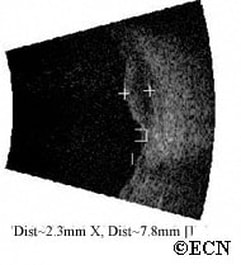
in a patient (female, 67 years old) with uveal melanoma. It originally was measured to be 2.6 mm in height and 8.7 mm in maximal diameter (Fig.1). If this tumor was located in the peripheral choroid, the optometrist would not have seen it. In our case, the patient refused treatment and after 1.5 years the tumor increased to11.9 in height (Fig.2). Enucleation with subsequent histopathologic evaluation revealed a large melanoma of predominantly epithelioid cell-type.
We have noticed that in all cases of rapid growth (of uveal melanoma) they were later determined to be of epithelial or mixed cell-types. In addition, some patients with spindle cell melanoma were observed to grow slowly (1 to 9 mm) over 14 years.
Best regards,
Prof. Alexander Bouiko
Ukrain
From: [email protected]
Date: January 30, 2005
I think most ophthalmic oncologists would say the tumor existed at the time the OD saw the patient. But, how much could it have grown in the internal of 11 months in diameter and in height?
I agree that the tumor was "likely" to have existed at the time of the OD's exam. But no one can prove it. Barrett Haik and Matt Wilson published a case - AJO - where a melanoma grew from nevus size to large in 6 months. I am sure you have had the experience of measuring tumor growth in 3 weeks, but would also agree that most small uveal melanomas will not demonstrate a measurable change in the same period.
Q: Could it have doubled in size?
A: Yes
Q: Tripled?
A: Yes
Q: Quadrupled?
A: Yes, but "could" is the operative word.
Q: By not identifying the tumor which provided to be a mixed cell malignant melanoma that was primarily made up of spindle cells and did not have vascular loops or networks was the patient a greater risk of metastasis than if the tumor eye was enucleated or treated with I-125 brachytherapy soon after the OD saw the patient?
A: I believe that if one did a multivariate analysis, largest tumor diameter would be the dominant factor.
The liver enzymes and imaging studies just prior to enucleation of the eye were all within normal limits. This is not uncommon and yet we all probably believe that there were micrometastases well before the ODs exam.
A: The timing of metastasis is a fascinating subject. If small melanomas have a 6% metastatic rate and large have a 38%, clearly waiting until a tumor is large will decrease a patients chance for survival.
The difficulty with these cases is that ODs who want all clinical privileges claim that they should not be held accountable because melanomas of the uveal tract are uncommon, they had little if any training in diagnosing such lesions.
They need to be held accountable, but by whom? The optometrists will not police themselves and would be unwilling to have ophthalmologists do it.
As optometrists assume clinical privileges and complications occur, the courts will play a greater role. I imagine that those who gave them clinical privileges will also be at legal risk. In this case, the patient either new he or she was getting a sub-optimal examination or was harmed by a false-sense that the exam was complete.
Warm regards,
Paul T. Finger, MD
New York City
DISCLAIMER: Postings on The ECN Mailing List are strictly the opinions of the authors. The ECN and its sponsors assume no responsibility for the accuracy of the information, nor do they assure the safety or effectiveness of any clinical recommendations in these postings.
Receive the latest news and opportunities from The Eye Cancer Foundation. Please fill out the form below.


