Small Juxtafoveal Melanoma Case 11
Tumor Board Discussion
A Case of a Small Juxtapapillary Melanoma
A Case of a Small Juxtapapillary Melanoma
|
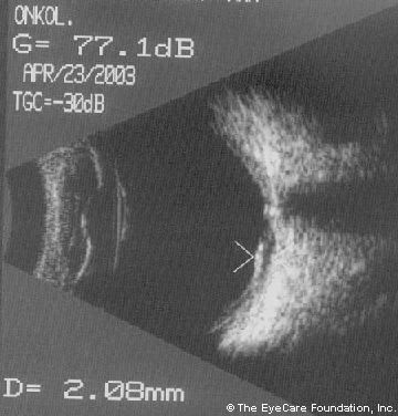
Ultrasound reveals a choroidal tumor with low internal reflectivity and an apical height of 2.1 mm
|
From: The Eye Cancer Network International Tumor Board
Date: Friday May 2nd, 2003 America/New_York To: ECN Group Subject: Case Reply-To: [email protected] Case Presentation: From: Grazyna Czechonska, MD ([email protected]) Date: Mon Apr 28, 2003 To: ECN International Tumor Board Subject: Case I present the case of a 39 year old graphic artist with no known systemic medical problems and a past ocular history of mild amblyopia OS. His vision has been noted to decline over a 2 month duration. I measured visual acuities of 20/20 OD and 20/30 OS three weeks ago. Within the last 3 weeks his vision has declined to 20/60 OS. He appears to have a small juxtafoveal choroidal melanoma. |
|
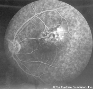
Fluorescein angiography reveals a small juxtafoveal tumor: approximately 7 x 5 mm.
Normally I would recommend 'close' observation for evidence of growth before an application of radioactive plaque.The problem is that he is a graphic artist. I want to save his vision. What do you think about TTT treatment alone in this case? Sandwich therapy? Plaque alone? Are there any other options for him in your opinion? Best regards, Grazyna Czechonska, MD Warsaw, Poland [email protected] |
Dgn. Malignant melanoma/ choroidal nevus
|
Responses to Dr. Czechonska:
From: Alexander Bouiko
Date: 5 May 2003 America/New_York
Subject: Re: The Eye Cancer Network - International Tumor Board
Dear Dr. Czechonska:
The present data of your patient is not quite demonstrative for melanoma. In such cases we prefer to perform antiinflammative, dehydratative treatment and to follow up the patient during 2-3 months. The clinic picture must be cleaner. If it will be the melanoma the preservation of VA will be very problematical.
Best regards,
Alexander Bouiko
From: Jaroslaw Kociecki
Date: 4 May 2003 America/New_York
Subject: Re: The Eye Cancer Network - International Tumor Board
Dear Dr. Czechonska,
I’d rather recommend close observation (especially with an aid of ultrasound) of this patient in order to see eventual progression. If the decreased vision is observed in course of secondary retinal detachment – it may be treated with gentle laser therapy or may sometimes disappear spontaneously (as I observed in some of our cases). If it really is a melanoma (and not a nevus) the patient will develop further deterioration of vision anyway - because of the tumor or because of eventual therapy. As to the latter one – I would recommend TTT which usually is quite effective in such small cases. You may expect significantly smaller post-treatment scotoma than after brachytherapy or “sandwich” therapy (which – in my opinion - is contraindicated in your case at least because of location and dimensions of the tumor). As an alternative you may consider proton beam irradiation (available in Germany), but this kind of treatment also produces similar scotomas, so the result may be very similar. As your patient is an artist, I’d recommend the close observation and if the progression develops – you may decide what kind of treatment will be most appropriate. In my experience with TTT – this kind of therapy is quite effective in such cases; nevertheless, the patient should realize his central vision will be probably irreversibly lost.
With kind regards,
Jaroslaw Kociecki
Poznan, Poland
From: Alain rousseau
Date: Thu, 1 May 2003 America/New_York
Subject: Re: The Eye Cancer Network - International Tumor Board
Dear Dr. Czechonska:
As for ttt, it depends how the tumor is located with regards to the fovea. I would favor waiting and watching closely, it will always be time to intervene, considering the risks involved. I am afraid TTT may cause substantial loss of vision because of its close relation with the disc. I have only little experience with ruthenium.
Alain Rousseau, MD
Quebec, Canada
From: Bertil Damato
Date: Thu, 1 May 2003
Subject: The Eye Cancer Network - International Tumor Board
Dear Dr. Czechonska,
It would be useful to see a colour photograph to determine whether or not orange pigment is present. In any case, the recent visual loss is suspicious of malignancy. I say to my patients with similar tumours that the conventional method of distinguishing a large naevus from a small melanoma is by observation, comparing the ophthalmoscopic appearances with baseline colour photographs and sequential ultrasonography. I mention that the chances of survival are related to the size of the tumour when it is treated and not how long it has been observed; however, I also point out that metastatic disease is incurable and mention that it is not known at what stage uveal melanomas start to disseminate, so that there is an uncertain risk involved in not treating a small melanoma immediately. At this point, some patients prefer to observe and others, with an identical tumor, feel happier with treatment, despite the visual loss. It is only fair to let the patient decide what risk to take.
With regards to treatment, my patients are given the choice between (1) TTT, as a simple and inexpensive outpatient procedure, but with about a 20% chance of failure, although this can be treated with radiotherapy at a later stage; (2) eccentric ruthenium plaque radiotherapy, without a posterior safety margin, if necessary, with adjunctive TTT at a later stage if the posterior tumour continues to extend; this has about a 98% chance of success in our series, but with a tumour so close to the fovea there is about a 70% chance of significant loss of central vision; (3) proton beam radiotherapy, which is more expensive and inconvenient, but which may result in less rapid loss of central vision than plaque radiotherapy, if loss of vision occurs.(In one or two patients like this the vision actually improved after proton beam radiotherapy, remaining good even after five years). Whatever type of radiotherapy is used, I would perform TTT without safety margins to improve vision by reducing the exudation from the tumour.
We have successfully treated a similar patient with endoresection combined with macular rotation, because he needed binocularity to continue to fly his plane. I would confirm that this patient actually does need binocularity, when balancing risks, because if he covers his eye with a patch or simply closes his eye he may find he can work reasonably well. He is a little fortunate in that the tumour is in the left eye so that any metamorphopsia or blurring is not as great a nuisance as it might have been if the tumour had developed in his right eye – and the patient might be comforted by this thought.
Yours sincerely,
Bertil Damato
Liverpool, UK
From: Ihab Saad Othman
Date: Wed, 30 Apr 2003
Subject: The Eye Cancer Network - International Tumor Board
Dear List Members:
As regard your notes, TTT is effective in small choroidal melanomas as primary treatment provided that the tumor is malignant and contain actively growing cells. Penetration of B rays (Ru-106) is low compared to Gamma rays (I-125). For a 2 mm thick tumor and an apical dose of 70 Gy, I-125 may be more hazardous to the fovea and optic disc. Chronic radiation damage of the retina is the result of a micro-occlusive vasculopathy. Thickening & hyaline degeneration in the vessel walls with areas of occlusion & concomitant loss of both capillary mural & endothelial cells with resultant obliterative endarteritis.The neural elements of the retina are more radioresistant than the vasculature. The changes are secondary to vascular occlusion with atrophy & thinning of the retina & atrophy of the ganglion cells. These changes appear late after brachytherapy (1.5-3 years) The incidence of optic nerve injury after plaque therapy is related to the dose, the dose rate , the size of the nuclide & the size and location of the tumour. An incidence of 9.8% complete optic atrophy & 3.8 % partial atrophy with a follow-up of 6-16 years was reported after Ruthenium 106 Plaques for posterior uveal melanoma. (Lommatzsch ,1986).
Sincerely,
Ihab Saad Othman, MD, FRCS
Consultant Ophthalmic Surgeon
Cairo University
From: Petr Soucek
Date: Wed, 30 Apr 2003
Subject: The Eye Cancer Network - International Tumor Board
Dear Dr. Czechonska,
Are color fundus photograph and ICG angiography available? As a treatment option I recommend our Gamma knife, which?is available closer to your city than any proton accelerator.
Best regards,
Petr Soucek, Prague
From: Wilko Friedrichs
Date: Tue Apr 29, 2003 4:43:18 PM America/New_York
Subject: The Eye Cancer Network - International Tumor Board
Dear Dr. Czechonska:
As an alternative to TTT I would consider Proton beam therapy with reduced safety margins at the foveal border of the tumor. Nevertheless VA might/will decrease considerably 2 to 4 years after treatment. For further details contact Prof. Foerster in Berlin, Dr. Chauvel in Nice/F.
Yours sincerely,
Wilko Friedrichs
Stuttgart, Germany
From: Alain rousseau
Date: Tue Apr 29, 2003 1:24:58 PM America/New_York
Subject: Re: The Eye Cancer Network - International Tumor Board
Dear Dr. Czechonska:
As for ttt, it depends how the tumor is located with regards to the fovea. I would favor waiting and watching closely, it will always be time to intervene, considering the risks involved.
Alain Rousseau, MD
Quebec, Canada
Dear Dr. Czechonska:
In this case I think the tumor should be considered as a suspicious naevus and not choroidal melanoma. If the visual loss is due to serous detachment of the macula I would recommend argon laser treatment of the surface of the naevus and close follow up.
Best regards,
Laurence Desjardins
Paris, France
DISCLAIMER: Postings on The ECN Mailing List are strictly the opinions of the authors. The ECN and its sponsors assume no responsibility for the accuracy of the information, nor do they assure the safety or effectiveness of any clinical recommendations in these postings.
Receive the latest news and opportunities from The Eye Cancer Foundation. Please fill out the form below.
Receive the latest news and opportunities from The Eye Cancer Foundation. Please fill out the form below.


