Slide Set Initiative
Internet-Based AJCC-UICC Pathology Slide Set
Team Leaders: Drs. Darryl Ainbinder, Tatyana Milman, Hans Grossniklaus, Sarah Coupland and Deepak Edward - Team Leaders
Eyelid Carcinoma Slide Set
Gigabyte high-resolution pathology imaging is now available. We are going to employ this new technology to teach ophthalmic pathology. Using the AJCC-UICC pTNM Classification System as a base, we will create a free, open-access ultra-high resolution teaching slide set. Both audio and video will be used to illustrate how the pTNM system was created to help diagnose (define) eyelid, conjunctival, iris, ciliary body, choroidal, retinal and orbital tumors.
Outline courtesy of Douglas Tkachuk, MD, FRCPC
President, Objective Pathology Ltd.
http://www.objectivepathology.com
How to Use Virtual Microscopy Tools
Instructional Videos: Example of instructional video featuring virtual microscopy.
Discussion Boards: Example of discussion board.
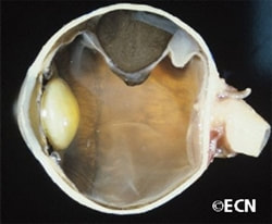
Examples of Virtual Microscopy (H&E Eyeballs, 20x Scans)
http://www.objectivepathology.com/Private/UHN/BrendaGallie/
Agenda
I. Collect case materials.
II. Select peer-reviewed representative examples.
III. Create case template (clinical history, contributor's name, metadata).
IV. Establish subgroup to create flash instructional videos.
V. Create password-protected or open-access discussion board.
VI. Establish open-access internet portals.
VII. Publish an AJCC-UICC print (DVD) version with Springer.
Learn about the caBig "SPIN" Shared Pathology Informatics Network together.


