Retinoblastoma Case 2- Tumor Board Presentation from Edward Averbukh, MD
|
Subject: Retinoblastoma Management Case Date: Wednesday March 7th, 2001
From: Edward Averbukh, MD Dear List Members: We have a difficult case and would like to get your comments on treatment. Case: A 5 y.o. girl with a past ocular history of bilateral retinoblastoma. One eye was enucleated. Other eye was treated (several times) with cryotherapy. She has received maximal possible chemotherapy |
|
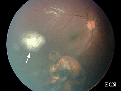
Pre: New small retinoblastoma temporal to the macula (arrow)
A new small lesion (see "pre" picture), appeared temporal to the macula. It is difficult to reach with cryo due to location and scaring. I treated the lesion by applying heavy green laser around the lesion and infrared laser - first time mostly on the lesion edges. There was no regression, in fact the lesion appeared more elevated.
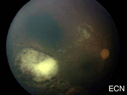
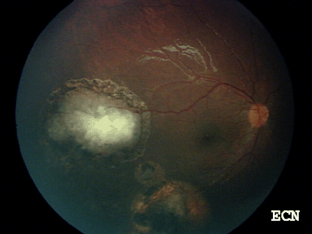
Then laser treatment was repeated twice - same technique (see "post" picture) - around the lesion green argon laser and on the lesion infrared diode (both through an indirect laser ophthalmoscope). However, the lesion looks much more elevated (now 4 weeks after a third treatment). That last treatment was really a heavy whitening application of both types of laser. On March 12, now 3 weeks after a very heavy laser treatment was applied to the tumor (infrared (diode) to the tumor itself and green (argon) around the lesion)
|
Image after treatment.
Question: What will be your approach:
Responses From ECN Members: "The tumor could be treated with either "cutting" cryo or more laser every 4 weeks. However, this is good place to use sub-tenon's carboplatin, with the laser. Since it appears to be somewhat recalcitrant to treatment, I would recommend this at the next EUA. If the tumor continues to not respond or threatens to extend beyond the scar now demarcating it, I think that a short course of chemotherapy with the current high dose and CSA would be worthwhile." |
Dear ECN Tumor Board:
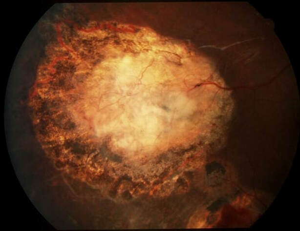
I am attaching here the last photograph of the lesion that was taken with regular fundus camera (not the Ret-cam, because there was no need for EUA).
To remind you, this monocular child was treated with full dose of chemotherapy, cryotherapy and brachytherapy (Ru-106) previously and the new lesion appeared on the edge of the previously treated area, so the scaring and the posterior location of the new lesion made the additional cryo difficult.
I applied laser through indirect laser ophthalmoscope. Periphery was treated with Argon and the lesion itself with IR (Iris Medical) laser, the total of 3 sessions few weeks apart. The lesion seemed to be growing rapidly following the laser treatment. However that was just swelling, probably due to inflammatory response, and 2 months later the tumor regressed.
Thank you all for your thoughts and advice in this difficult case.
DISCLAIMER: Postings on The ECN Mailing List are strictly the opinions of the authors. The ECN and its sponsors assume no responsibility for the accuracy of the information, nor do they assure the safety or effectiveness of any clinical recommendations in these postings.
Receive the latest news and opportunities from The Eye Cancer Foundation. Please fill out the form below.
Receive the latest news and opportunities from The Eye Cancer Foundation. Please fill out the form below.

