Large Intraocular Tumor Case 12
Tumor Board Presentation from Paul T. Finger, MD and Steven A. McCormick, MD
Subject: Unknown
Date: September 17th, 2003
Dear List Members
We have a interesting case and would like to get your comments, particularly the ophthalmic pathologists in our group.
Case
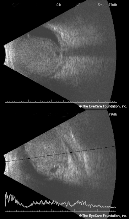
This 38 year old female noted 8 months of progressive loss of vision OD, then a sudden painful proptosis which progressed over 7 days. Ophthalmic examination revealed neovascular glaucoma, a restricted globe and 8 mm of axial proptosis. There was no view of the fundus so ultrasonography was performed.
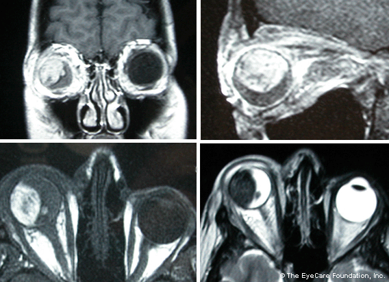
Only the orbits were positive. This scan was significant for an intraocular tumor which displays high signal intensity on T1 weighted images. On T2 weighted images it displayed low signal intensity. (These findings can be seen in melanoma and hemorrhage). Though scleritis was noted, there was no evidence of extraocular tumor extension.
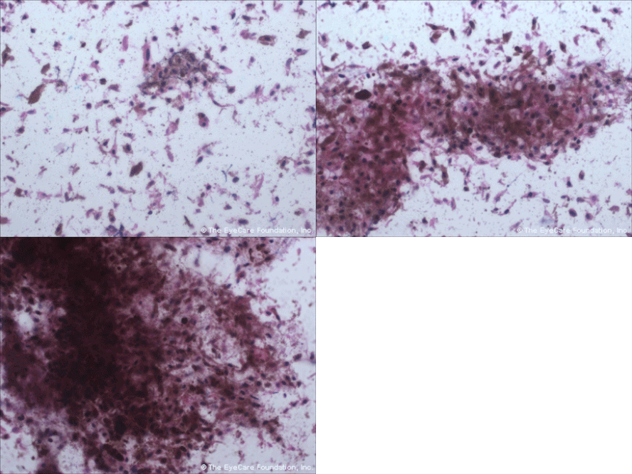
We discussed the relative risks and benefits of primary enucleation versus fine-needle aspiration biopsy with possible enucleation based on cytology. The results are available below.
|
Within days this patient responded dramatically to steroids, antibiotics and glaucoma medications. She is currently NLP and comfortable, with no orbital signs.
What will be your approach in this case? 1. What is your presumptive diagnosis? 2. What ancillary tests would you order? 3. Is another biopsy recommended? 4. Suggested treatments? Final Diagnosis Necrotic Malignant Choroidal Melanocytoma DISCLAIMER: Postings on The ECN Mailing List are strictly the opinions of the authors. The ECN and its sponsors assume no responsibility for the accuracy of the information, nor do they assure the safety or effectiveness of any clinical recommendations in these postings. |
Fine Needle Aspiration Biopsy Cytology
|
Receive the latest news and opportunities from The Eye Cancer Foundation. Please fill out the form below.


