Bilateral Angiomatosis of the Iris
Therapeutic Challenge
History: A 65-year old white female was sent to the New York Eye Cancer Center with a history of recurrent spontaneous hyphemas in her left eye. They started 2.5 years ago and she has had 4 episodes.
Ophthalmic Examination:
Visual acuity was measured to be 20/16 in the right eye and 20/20 in the left. Intraocular pressures by tonometry were 16 mm/hg in the right eye and 17 in the left.
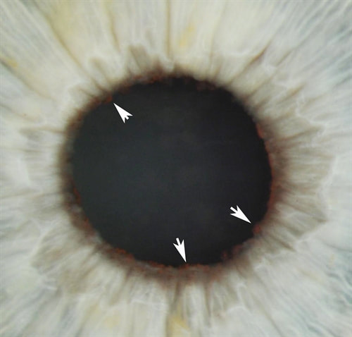
Anterior segment examination revealed numerous small vascular tumors at the pupillary margin in the left iris more than the right undefined.
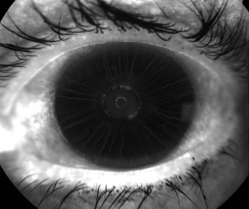
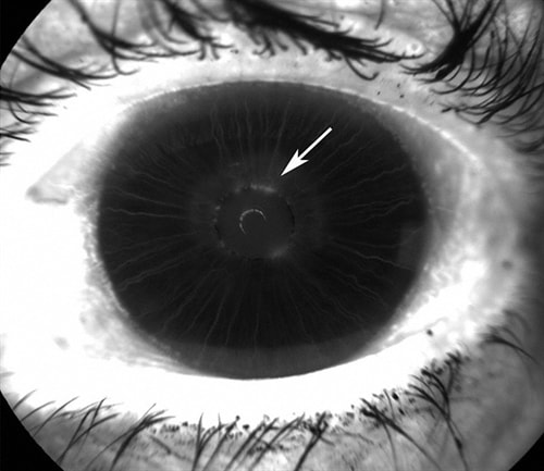
Similarly, Top - fluorescein angiography revealed early hyperfluorescence from the tumors in the left eye with late leakage of dye into the anterior chamber - Bottom image.
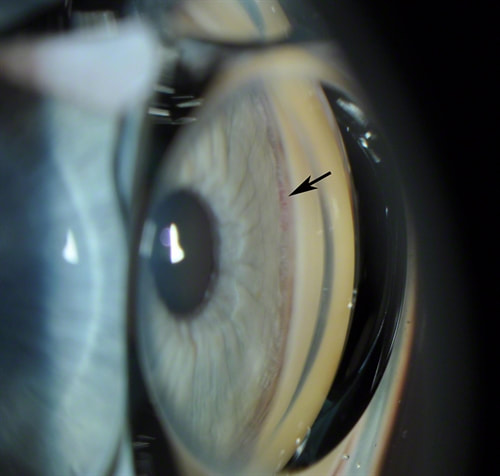
Gonioscopy showed small blood vessels, bridging the angle in the temporal quadrant in the left eye undefined.
High frequency ultrasound imaging (UBM) revealed no evidence of tumor (benign or malignant) upon examination of 360-degree anterior segment of the left eye. Maximal iris thickness was 0.7 mm, maximum ciliary body thickness was 1.5 mm. and pars plana cysts were seen in the nasal quadrant. The right eye was unremarkable.
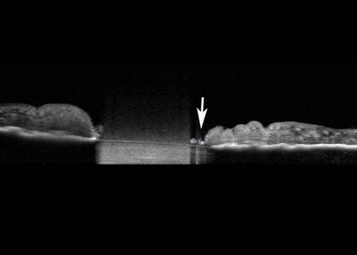
Anterior Segment Optical Coherence Tomography: Nodular lesions seen at pupillary margin. Hemorrhage seed as opacities in the anterior chamber undefined
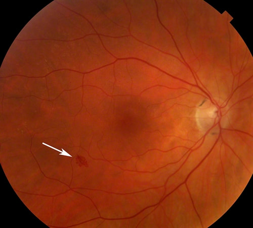
Bilateral indirect ophthalmoscopic examination was significant for a small intraretinal hemorrhage in the right macula and internal limiting membrane wrinkling in he left. No retinal neovascularization was noted. undefined
A hematology oncology consultation was obtained:
-CBC: Mild decrease in white blood cell count (3.0x10³/ul; normal = 3.6-11.2x10³ /ul)
-PT/PTT/bleeding time – within normal range
-Serum glucose level (103 mg/dl; normal range 65-99 mg/dl)
-Serum alkaline phosphatase (39 IU/L; normal range 47-112 IU/L)
-Serum protein electrophoresis normal
-Computed tomography of the brain, chest abdomen and pelvis normal
Further work-up or suggested Treatment?
Receive the latest news and opportunities from The Eye Cancer Foundation. Please fill out the form below.

